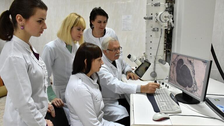
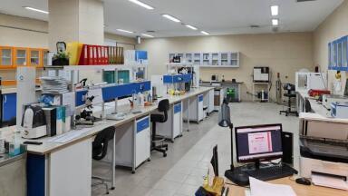
Environmental laboratory
Instead of recording only image intensity at each point, these approaches capture additional information across space, energy, reciprocal space and time.
The result is a much richer dataset that links structure, chemistry and behaviour within the same experiment.
Traditional TEM or STEM typically produces a 2D projection of a 3D object, with contrast that can be difficult to interpret unambiguously. Multidimensional methods address this by adding new measurement axes.
Electron tomography reconstructs full three-dimensional structures by combining images taken at multiple tilt angles.
Spectroscopic techniques such as EDS and EELS add an energy dimension, allowing elemental composition and chemical state to be mapped alongside structure. 4D-STEM goes further by recording a full diffraction pattern at every scan position, combining real-space and reciprocal-space information to quantify strain, crystallography and internal fields with high precision.
A further dimension is time. In-situ and time-resolved electron microscopy uses specialised holders and detectors to observe samples while they are heated, electrically biased, mechanically stressed or exposed to reactive gases or liquids.
This makes it possible to watch transformations as they occur, rather than inferring mechanisms from before-and-after images. Increasingly, these dimensions are combined within single experiments, supported by high-speed cameras and data-intensive computational analysis.
The relevance of multidimensional electron microscopy to environmental monitoring lies in its ability to reveal mechanisms that are invisible to field instruments. Typical analytical systems measure bulk properties such as concentration, mass or optical response.
Multidimensional electron microscopy explains what those signals actually represent at the particle or interface level, and how they change over time.
By resolving structure, chemistry, and dynamics together, the technology provides definitive evidence about contaminant form, reactivity and stability. This supports better sensor design, more defensible proxy measurements and stronger links between monitoring data and environmental risk.
For monitoring professionals, it functions as a reference tool that underpins interpretation, validation and forward-looking environmental management rather than a replacement for routine monitoring itself.
In air quality monitoring, the most critical uncertainties often relate to particulate matter rather than bulk gas concentrations. Multidimensional electron microscopy allows individual particles to be analysed in terms of structure, composition and chemical state.
Techniques such as EELS and advanced STEM reveal oxidation states and elemental associations that determine toxicity and atmospheric reactivity.
Time-resolved and in-situ experiments make it possible to observe how particles oxidise or hydrate after emission, helping explain gaps between emission inventories and ambient measurements.
These insights support air monitoring by improving source attribution, refining sensor calibration and strengthening links between measured particle metrics and health impacts.
Water monitoring frequently encounters contaminants that are mobile, transformed or associated with particles rather than present as simple dissolved species.
Multidimensional electron microscopy enables direct characterisation of nanoparticles, colloids and other sediment-bound materials that govern contaminant transport and bioavailability.
By combining spatial and energy dimensions, it becomes possible to identify how pollutants partition between mineral phases and organic matter, particularly at sediment–water interfaces.
In-situ techniques further reveal how these systems respond to changes in, for instance, pH or salinity conditions, informing both monitoring strategies and the evaluation of treatment performance.
In soils, key environmental processes occur at the scale of pores and organo-mineral complexes. Electron tomography and spectroscopic mapping allow soil microstructure to be reconstructed in three dimensions and chemical forms of nutrients or contaminants to be identified.
This helps explain why some pollutants remain immobilised while others migrate into groundwater or become bioavailable.
Time-resolved microscopy also supports controlled observation of weathering and degradation processes, providing mechanistic evidence that complements long-term field monitoring data and improves confidence in risk assessments.
Across all environmental applications, multidimensional electron microscopy provides ground-truth evidence at the smallest relevant scales.
It explains the mechanisms behind signals detected by sensors and sampling networks, turning environmental monitoring from a largely descriptive activity into a predictive, process-based discipline.
This capability is increasingly important as monitoring frameworks shift toward source attribution and risk-based thresholds.
IET Guide 2026

 Product directory














.jpg)