Laboratory microscope
Materials destined for space exploration face conditions far more extreme than those found in aviation or terrestrial applications. Spacecraft components endure temperature swings from over +150°C in direct sunlight to below –150°C in shadow along with ionising radiation, vacuum and atomic oxygen in low Earth orbit. These factors accelerate degradation in metals, composites and polymer-based materials and create major challenges for long-term reliability. Here, Dr Herman Lemmens, Sales Development Director EMEA at Thermo Fisher Scientific explains how advanced microscopy helps researchers evaluate and strengthen materials for space missions.
Ensuring the structural integrity and long-term functionality of space materials relies on detailed insight into microstructure, chemistry and defect evolution. Conventional bulk testing cannot fully predict how materials behave under such extreme conditions. Moreover, many failure mechanisms begin at length scales far smaller than traditional mechanical tests can detect, making high-resolution imaging essential for accurate lifetime prediction. Advanced microscopy techniques that probe from the microscale to the nanoscale have therefore become indispensable for qualification and design.
Space environments introduce degradation pathways that rarely appear on Earth. Metallic alloys may develop radiation-induced defect clusters, grain boundary embrittlement or voids. Composites can delaminate under repeated thermal cycling as layers expand and contract at different rates, while thermal protection systems on re-entry vehicles experience ablation and oxidation and polymer surfaces in low Earth orbit erode under exposure to atomic oxygen. Also, optical components, such as mirrors and sensor housings, can suffer darkening or micro-pitting that degrades performance over time.
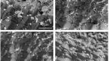
Microscopy allows engineers to directly observe these processes. Scanning electron microscopy provides high-resolution surface imaging to reveal corrosion pits, microcracks and fracture patterns. When paired with energy-dispersive X-ray spectroscopy it can map elemental distributions and identify chemical variations that influence durability.
Transmission electron microscopy provides insight at the atomic and nanoscale, resolving dislocation networks, irradiation-induced voids and nanoscale precipitates. These features often dictate mechanical properties and failure modes, making nanoscale visibility essential for linking microscopic defects to macroscopic performance. Cryo-TEM workflows are increasingly being explored for analysing polymers and composite matrices, preserving radiation-induced structural changes that would otherwise be altered during preparation.
Three-dimensional approaches such as focused ion beam scanning electron microscopy allow reconstruction of subsurface structures, including crack paths and porosity networks. This volumetric capability is particularly valuable for fatigue assessment in alloys and composites, where internal defects may compromise long-term structural integrity.
By uniting surface imaging, chemical mapping and volumetric reconstruction, microscopy enables a comprehensive understanding of how materials respond to radiation, vacuum and thermal extremes. These insights support predictive models and guide preventive engineering strategies. Such datasets increasingly feed into machine-learning models that simulate long-term degradation, helping engineers reduce the number of physical life-cycle tests required.
Additive manufacturing (AM) is increasingly used in aerospace for its ability to produce lightweight lattice structures, consolidated geometries and complex forms that cannot be made conventionally. For spaceflight, the potential for in-orbit production adds further value. Yet AM introduces microstructural complexities that must be quantified before flight qualification. Porosity, unmelted powder particles and anisotropic grain growth can form weak points that reduce fatigue life.
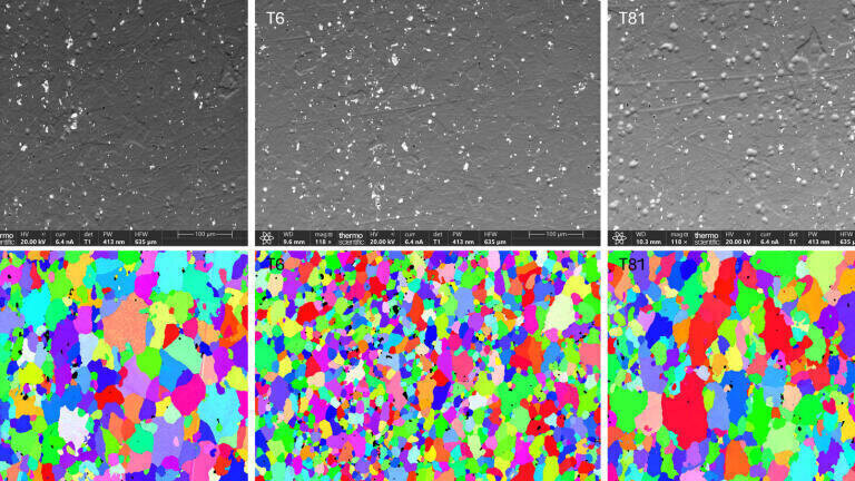
Microscopy provides a rigorous framework for assessing AM materials. Correlative workflows that combine scanning electron microscopy (SEM), energy-dispersive X-ray spectroscopy (EDS) and electron backscatter diffraction (EBSD) allow engineers to evaluate surface quality, chemical composition and crystallographic texture within the same region. EDS identifies inclusions or unmelted particles, whereas EBSD maps grain orientations that influence anisotropic mechanical behaviour and can reveal build-related structural signatures.
These multi-scale and multi-modal insights support deeper understanding of the relationship between AM process parameters and final material performance. By identifying regions susceptible to crack initiation or fatigue, engineers can refine build conditions, heat treatments or post-processing steps to increase reliability without relying solely on destructive testing. As AM moves toward certification for structural flight components, microscopy-based quality protocols are becoming an essential part of standardisation across the aerospace supply chain.
Protective coatings are central to spacecraft design, serving as thermal barriers, erosion-resistant layers or radiation-shielding films. Their performance depends not only on bulk chemistry but also on microstructural details such as porosity, adhesion between layers and phase distribution.
Microscopy offers a direct method to quantify and optimise these properties. Cross-sectional SEM with EDS can reveal how stabilising elements such as yttrium, zirconium or magnesium are distributed in ceramic thermal barrier coatings. Uniform distribution supports improved oxidation resistance and thermal stability.
EBSD contributes further insight into crystallographic orientation and phase differences within and across coating layers. These maps can identify mismatches that concentrate stress and lead to spallation during repeated thermal cycling. By correlating microstructural observations with environmental testing, engineers can refine deposition parameters for plasma spraying, chemical vapor deposition and emerging additive coating methods. This iterative approach strengthens adhesion, reduces thermal fatigue and extends component lifetime.
Advanced microscopy offers multi-scale visibility that bridges nanoscale defects observed in transmission electron microscopy with microscale surface features seen in scanning electron microscopy. Three-dimensional characterisation through focused ion beam serial sectioning permits volumetric reconstruction of internal porosity, crack networks and hidden defects. Correlative analysis that integrates structural imaging, chemical mapping and crystallography allows engineers to link microstructural features to performance in a direct and predictive way.
Microscopy also enables early identification of potential failure modes, supporting proactive design changes and process optimisation. Its versatility across metals, composites, polymers, coatings and additively manufactured parts allows engineers to address the full range of space materials challenges. Together these capabilities make microscopy a predictive platform for innovation and a vital tool in the development of lighter, stronger and longer-lasting spacecraft components.
Advanced microscopy has become central to the development and qualification of materials for space exploration. By revealing degradation mechanisms, validating additive manufacturing quality and optimising coating performance, techniques such as SEM, EDS and EBSD allow engineers to connect microstructural understanding with design decisions. In an environment where reliability is paramount and failure is costly, these imaging and analysis tools support informed choices and drive the development of robust materials for next-generation missions.
IET 36.2 Mar/Apr 2026


.jpg)